Nervous tissue is a specialized type of tissue found in the bodies of animals, including humans. It is responsible for the transmission, integration, and processing of electrical signals throughout the body. Nervous tissue forms the basis of the nervous system, which includes the brain, spinal cord, and peripheral nerves.
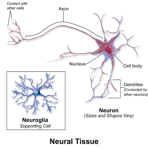
Nervous tissue is composed of two main types of cells: neurons and neuroglia (also known as glial cells). Neurons are the functional units of the nervous system and are specialized for transmitting electrical impulses. They consist of a cell body (soma), dendrites that receive signals from other neurons or sensory receptors, and an elongated axon that transmits signals to other neurons, muscles, or glands.
Overall, nervous tissue, comprised of neurons and neuroglia, forms the foundation of the intricate nervous system. It enables the transmission of electrical signals, facilitates communication between various parts of the body, and plays a vital role in regulating bodily functions and responses.
Definition of Nervous tissue
Nervous tissue, also known as neural tissue, is a specialized type of tissue found in the nervous system. It is responsible for transmitting and processing electrical signals, allowing for communication and coordination within the body. Nervous tissue is composed of two main types of cells: neurons and neuroglia (glial cells).
Location of Nervous Tissue
Nervous tissue is located in various parts of the body, primarily in the central nervous system (CNS) and peripheral nervous system (PNS).
In the CNS, nervous tissue is extensively present in the brain and spinal cord. The brain, located in the cranial cavity, is responsible for higher cognitive functions, sensory processing, motor coordination, and the regulation of bodily functions. It consists of different regions, including the cerebral cortex, cerebellum, brainstem, and subcortical structures, all of which contain dense networks of nervous tissue. The spinal cord, enclosed within the vertebral column, acts as a pathway for transmitting signals between the brain and the rest of the body.
The PNS comprises the branching peripheral nerves that extend throughout the body. These nerves connect the CNS to the sensory organs, muscles, glands, and other organs. Nervous tissue within the PNS enables communication and control over bodily functions. The peripheral nervous system (PNS) is further divided into two main components: the somatic nervous system and the autonomic nervous system. The somatic nervous system regulates voluntary movements and sensory perception, while the autonomic nervous system controls involuntary functions like heartbeat, digestion, and respiration.
Within the nervous tissue, the key components are neurons, also known as nerve cells. Neurons are the functional units of the nervous system, responsible for transmitting electrical signals called action potentials. These impulses are generated in response to stimuli and travel along the length of the neuron's axon to communicate with other neurons, muscles, or glands.
Therefore, nervous tissue can be found in the peripheral nerves throughout the body, as well as in the organs of the central nervous system, such as the brain and spinal cord. It is within these locations that neurons carry out their functions of receiving,transmitting information,and integrating, enabling the complex functioning of the nervous system.
Characteristics Of Nervous Tissue
Nervous tissue possesses several distinct characteristics that contribute to its specialized functions in the nervous system. Here are the key characteristics of nervous tissue:
1. Excitability: Nervous tissue is highly excitable, meaning it can respond to stimuli and generate electrical signals called action potentials. This characteristic allows for the rapid transmission of information within the nervous system.
2. Conductivity: Nervous tissue has the ability to conduct electrical impulses over long distances. This is achieved through the propagation of action potentials along the length of neurons, allowing for the transmission of signals from one part of the body to another.
3. Cell-to-Cell Communication: Nervous tissue facilitates rapid and precise communication between cells. Neurons are specialized cells that transmit signals to other neurons or target cells, such as muscles or glands, through synapses. This allows for the coordinated functioning of different parts of the body.
4. High Metabolic Rate: Nervous tissue has a high metabolic rate, requiring a constant supply of oxygen and glucose to meet its energy demands. The brain, in particular, is highly metabolically active and consumes a significant amount of energy.
5. Complexity and Specialization: Nervous tissue is highly complex and specialized. Neurons have unique structures, such as dendrites that receive incoming signals and axons that transmit signals to other cells. Different types of neurons have distinct shapes and functions based on their roles in sensory perception, motor control, or information processing.
6. Plasticity: Nervous tissue exhibits plasticity, which refers to its ability to change and adapt in response to internal and external stimuli. This allows for learning, memory formation, and the ability to reorganize neural connections in response to experiences or injuries.
7. Neuroglia Support: Nervous tissue is supported by specialized cells called neuroglia or glial cells. These cells provide structural support, insulation, and nutrition to neurons. They also play roles in immune defense, the removal of waste products, and the maintenance of the extracellular environment.
8. Integration and Processing of Information: Nervous tissue is responsible for the integration and processing of sensory information, as well as the generation of appropriate responses. This includes sensory perception, motor control, memory, and cognition.
9. Neurotransmitters: Neurotransmitters are chemical messengers that play a crucial role in the transmission of signals within the nervous system. They are released by neurons and facilitate communication between neurons, as well as between neurons and their target cells, such as muscles or glands. Neurotransmitters are stored in vesicles within the presynaptic neuron and are released into the synapse, the small gap between the presynaptic neuron and the postsynaptic neuron or target cell.
10. Synapses: Synapses are specialized junctions that allow for communication between neurons and the transmission of signals within the nervous system. They are the points of contact between the presynaptic neuron (sending neuron) and the postsynaptic neuron (receiving neuron) or target cell.
Overall, the characteristics of nervous tissue allow for the complex and precise functioning of the nervous system, enabling the coordination of various physiological processes and behaviors in the body.Nervous tissue is a vital component of the nervous system, consisting of neurons and glial cells. It is characterized by specialized structures such as dendrites and axons, which facilitate the transmission of signals. Neurotransmitters play a key role in the communication between neurons, which occurs at synapses. Nervous tissue exhibits longevity, although regenerative capacity varies among different types of neurons. These characteristics collectively enable nervous tissue to coordinate, communicate, and regulate the various activities of the body within the complex network of the nervous system.
Structure of Nervous tissue
Nervous tissue is composed of two main types of cells: neurons and neuroglia (or glial cells). Here is a breakdown of their structures:
1. Neurons:
Cell Body (Soma): The main part of the neuron containing the nucleus and other cellular organelles. It is responsible for maintaining the metabolic functions of the cell.
Dendrites: Branch-like extensions that receive signals from other neurons or sensory receptors. They increase the surface area for synaptic connections.
Axon: A long, slender projection that transmits signals away from the cell body. It is covered by a myelin sheath in some neurons, which insulates and speeds up the conduction of electrical impulses.
Axon Terminal: The end of the axon that forms synapses with other neurons or target cells. Neurotransmitters are released from the axon terminals to transmit signals across synapses.
2. Neuroglia (Glial Cells):
Astrocytes: Provide structural support to neurons, regulate the chemical environment of the brain, and facilitate nutrient and waste exchange between neurons and blood vessels.
Oligodendrocytes (in CNS) and Schwann Cells (in PNS): Form myelin sheaths around axons, which increase the speed and efficiency of signal transmission.
Microglia: Act as immune cells in the central nervous system, defending against pathogens and removing cellular debris.
Ependymal Cells: Line the ventricles of the brain and the central canal of the spinal cord. They help produce cerebrospinal fluid and assist in its circulation.
Additionally, nervous tissue contains blood vessels that provide oxygen, nutrients, and remove waste products from the cells.
The overall structure of nervous tissue involves the organization of neurons and glial cells into complex networks and circuits. These networks allow for the transmission, processing, and integration of electrical and chemical signals, which underlie the functioning of the nervous system and the control of bodily activities.
Types of Nervous tissue
1.
Neurons
2. Neuroglia
1. Neurons
There are several different types of neurons, each with specific functions and characteristics. Here are some of the main types of neurons:
1. Sensory Neurons: Sensory neurons, also known as afferent neurons, are responsible for transmitting sensory information from sensory organs (such as the eyes, ears, skin, etc.) to the central nervous system (CNS). They detect external stimuli and convert them into electrical signals that can be interpreted by the brain.
2. Motor Neurons: Motor neurons, also called efferent neurons, transmit signals from the CNS to muscles, organs, or glands. They are involved in controlling and coordinating voluntary and involuntary movements and responses. Motor neurons initiate muscle contractions and regulate various bodily functions.
3. Interneurons: Interneurons, also known as association neurons or local circuit neurons, are located entirely within the CNS. They act as intermediaries between sensory and motor neurons, relaying and integrating signals within the CNS. Interneurons play a crucial role in processing and interpreting information, making decisions, and coordinating complex functions.
4. Mirror Neurons: Mirror neurons are a specialized type of neurons that are active both when an individual performs a specific action and when they observe someone else performing the same action. They are believed to be involved in social cognition, empathy, and understanding the intentions and actions of others.
5. Relay Neurons: Relay neurons, also called projection neurons or relay cells, are responsible for transmitting signals between different regions of the CNS. They connect different areas of the brain and spinal cord, allowing for communication and integration of information across various brain regions.
These are just a few examples of the diverse types of neurons found in the nervous system. Each type plays a specific role in transmitting and processing information, allowing for the complex functions of the nervous system. It's important to note that these types of neurons can vary in structure and function across different species and within specific regions of the nervous system.
Neuroglia, also known as glial cells or glia, are a group of non-neuronal cells that provide support and protection to neurons in the nervous system. While neurons are the primary cells responsible for transmitting electrical signals, neuroglia play crucial roles in maintaining the structural integrity of nervous tissue, regulating the chemical environment, and supporting the functions of neurons. There are several types of neuroglia
:
 |
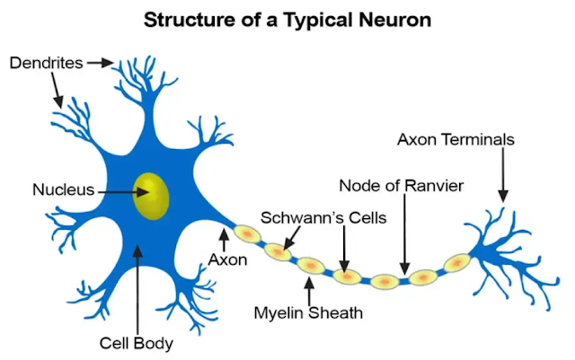
| Labeled diagram of a neuron (neural tissue). |
Structure of Neuron/Parts of a neuron
A neuron, also known as a nerve cell, is the fundamental unit of the nervous system. It has several distinct parts that contribute to its structure and function. The main parts of a neuron are as follows:
1. Cell Body (Soma): The cell body is the main portion of the neuron that contains the nucleus, as well as other organelles responsible for the cell's metabolic functions. It integrates incoming signals from dendrites and generates outgoing signals through the axon.
2. Dendrites: Dendrites are the branched extensions that extend from the cell body. They receive incoming signals from other neurons or sensory receptors and transmit those signals to the cell body. Dendrites play a crucial role in the integration of synaptic inputs.
3. Axon: The axon is a long, slender projection that arises from the cell body. It conducts electrical signals, known as action potentials, away from the cell body toward other neurons or target cells. The axon can vary in length, ranging from a fraction of a millimeter to several feet long.
4. Axon Hillock: The axon hillock is the cone-shaped region of the neuron located between the cell body and the axon. It plays a critical role in the initiation of action potentials by integrating and triggering the generation of electrical signals.
5. Myelin Sheath: The myelin sheath is a fatty substance that surrounds and insulates some axons, allowing for faster transmission of electrical signals. It is formed by specialized glial cells called oligodendrocytes in the central nervous system and Schwann cells in the peripheral nervous system.
6. Nodes of Ranvier: Nodes of Ranvier are small gaps or interruptions in the myelin sheath along the axon. They facilitate the saltatory conduction of action potentials, allowing the electrical signals to "jump" from one node to the next, which speeds up the signal transmission.
7. Axon Terminals: At the end of the axon, there are specialized structures called axon terminals or synaptic terminals. These terminals form synapses with other neurons or target cells. They contain synaptic vesicles that store neurotransmitters, which are released into the synapse to transmit signals to the next neuron or target cell.
These parts of a neuron work together to receive, integrate, and transmit electrical signals, enabling communication and coordination within the nervous system. Each part has a specific function that contributes to the overall functioning of the neuron and the nervous system as a whole.
Shapes of neuron
Neurons can exhibit various shapes or morphologies, reflecting their specific functions and locations within the nervous system.
Here are some common shapes or classifications of neurons:
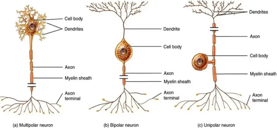
1. Multipolar Neurons: Multipolar neurons are the most common type of neurons. They have multiple processes or extensions emanating from the cell body. One of these extensions is the axon, while the rest are dendrites. The cell body is typically located off to the side, and the dendrites receive incoming signals from other neurons. Multipolar neurons are involved in a wide range of functions and are found throughout the central nervous system.
2. Bipolar Neurons:Bipolar neurons have two processes extending from the cell body—an axon and a dendrite. One end of the neuron has the dendrite, which receives incoming signals, and the other end has the axon, which transmits signals away from the cell body. Bipolar neurons are commonly found in specialized sensory organs, such as the retina of the eye and the olfactory epithelium in the nasal cavity.
3. Unipolar (Pseudounipolar) Neurons: Unipolar neurons have a single extension that splits into two branches, resembling a T-shape. This single process acts as both an axon and a dendrite. The cell body is typically located to the side. Unipolar neurons are commonly found in the peripheral nervous system and are involved in transmitting sensory information.
4. Anaxonic Neurons: Anaxonic neurons are characterized by having no apparent axon. Instead, they have multiple dendrites that receive and integrate signals. Anaxonic neurons are often found in the brain and are involved in processes such as visual perception and information processing. Their function is less understood compared to other neuron types.
These classifications describe general morphological characteristics of neurons, but it's important to note that the actual structure of neurons can vary widely, and there may be subtypes or variations of these general shapes depending on the specific functions and locations within the nervous system. Additionally, some neurons can exhibit a combination of features from different types, further adding to the complexity and diversity of neuronal structures.
Types of neuron
Neurons, the fundamental cells of the nervous system, can be categorized into different types based on their structure and function.
I. Types of Neurons Based on Structure
:
 |
| Three shapes of neurons |
1. Unipolar Neurons: Unipolar neurons have a single process extending from the cell body. This process acts as both an axon and a dendrite. Unipolar neurons are commonly found in the peripheral nervous system (PNS) and serve as sensory neurons, relaying sensory impulses from various parts of the body to the central nervous system (CNS).
2. Bipolar Neurons: Bipolar neurons have two processes that extend from opposite ends of the cell body: one dendrite and one axon. These neurons are relatively less common and are primarily found in specialized sensory organs such as the retina of the eye, the cochlea of the ear, and the olfactory epithelium (responsible for detecting smell).
3. Multipolar Neurons: Multipolar neurons possess multiple processes emanating from the cell body, consisting of numerous dendrites and a single axon. Most neurons in the CNS are multipolar neurons. They play various roles in the integration and processing of information within the brain and spinal cord.
II. Types of Neurons Based on Function:
1. Sensory Neurons (Afferent Neurons): Sensory neurons carry sensory impulses from the sensory receptors (e.g., skin, muscles, organs) to the CNS. They provide information about touch, pain, temperature, pressure, and proprioception (awareness of body position).
2. Motor Neurons (Efferent Neurons): Motor neurons transmit motor impulses from the CNS to the effectors, such as muscles and glands, enabling voluntary and involuntary movements and actions.
3. Interneurons (Association Neurons): Interneurons are found entirely within the CNS and serve as connectors between sensory and motor neurons. They integrate and process information, facilitating communication within the CNS and enabling complex neural functions.
4.Pyramidal Neurons: Pyramidal neurons are a type of excitatory neuron found in the cerebral cortex, particularly in areas responsible for higher cognitive functions. They have a distinct pyramid-shaped cell body and play a crucial role in processes like learning, memory, and decision-making.
5.Purkinje Cells: Purkinje cells are large, specialized neurons located in the cerebellum, a part of the brain responsible for motor coordination and balance. They receive inputs from various sources and are involved in fine-tuning and modulating motor activities.
6.Bipolar Neurons: Bipolar neurons have two distinct processes or extensions emerging from the cell body—one axon and one dendrite. They are commonly found in specialized sensory organs like the retina of the eye and the olfactory epithelium in the nose. Bipolar neurons help in relaying sensory information to other neurons.
These different types of neurons, based on their structure and function, work together to enable the complex functions of the nervous system, including sensory perception, motor control, cognitive processes, and the regulation of bodily functions.
2. Neuroglia
Neuroglia, also known as glial cells or simply glia, are a type of non-neuronal cells that provide support and protection to neurons in the nervous system. While neurons are the primary cells involved in transmitting and processing information, glial cells play crucial roles in maintaining the structural integrity of the nervous system and supporting neuronal functions
.
There are several types of glial cells, each with its own functions and characteristics. The main types of glial cells are:
1. Astrocytes: Astrocytes are the most abundant and diverse type of glial cells in the central nervous system (CNS). They have numerous functions, including providing structural support to neurons, regulating the concentration of ions and neurotransmitters in the extracellular space, forming the blood-brain barrier, and participating in the repair process after neuronal injury.
2. Oligodendrocytes: Oligodendrocytes are specialized glial cells found in the CNS. Their primary function is to produce and maintain myelin, a fatty substance that forms a protective sheath around axons, which enhances the speed and efficiency of nerve impulse transmission.
3. Schwann Cells: Schwann cells are the glial cells found in the peripheral nervous system (PNS). Similar to oligodendrocytes, they are responsible for producing myelin. Schwann cells wrap around peripheral nerve axons, forming myelin sheaths that facilitate rapid conduction of nerve impulses.
4. Microglia: Microglia are the resident immune cells of the CNS. They act as the primary defense mechanism against pathogens and foreign substances in the brain. Microglia are involved in immune responses, phagocytosis (engulfing and removing cellular debris), and modulating inflammation in the CNS.
5. Ependymal Cells: Ependymal cells are specialized glial cells that line the ventricles of the brain and the central canal of the spinal cord. They have ciliated surfaces and are involved in the production and circulation of cerebrospinal fluid (CSF), which provides cushioning and support to the brain and spinal cord.
These different types of glial cells work together to maintain the overall function and health of the nervous system. While historically considered supporting cells, research has revealed that glial cells have active roles in regulating neuronal communication, synaptic function, and neural development. The study of glial cells, known as glial biology or neuroglia research, has gained increasing attention in recent years for its significance in understanding brain function and neurological disorders.
Function Of Nervous Tissue
The nervous tissue has several important functions in the body:
1. Communication: Nervous tissue allows for the rapid transmission of signals and information throughout the body. It facilitates communication between different cells, tissues, and organs, enabling coordination and integration of various physiological processes.
2. Sensory Reception: Nervous tissue is responsible for detecting and responding to stimuli from the external and internal environment. Specialized sensory receptors convert sensory stimuli such as light, sound, touch, temperature, and chemical signals into electrical impulses that can be processed and interpreted by the nervous system.
3. Integration and Processing: Nervous tissue processes and integrates sensory information. It analyzes and interprets incoming signals, combining them with stored information, memories, and learned behaviors. This integration allows for the generation of appropriate responses and the coordination of bodily functions.
4. Motor Control: Nervous tissue controls and coordinates voluntary and involuntary movements. It generates signals that travel from the central nervous system to muscles and glands, initiating and regulating muscle contractions and glandular secretions.
5. Regulation of Homeostasis: Nervous tissue plays a vital role in maintaining homeostasis, the stable internal environment necessary for optimal bodily function. It regulates physiological processes such as heart rate, blood pressure, body temperature, respiration, digestion, and hormone secretion to ensure a balance between different systems.
6. Memory and Learning: Nervous tissue is involved in memory formation and learning processes. It enables the storage, retrieval, and consolidation of information, allowing for the acquisition of knowledge, skills, and experiences.
7. Emotional Responses: Nervous tissue contributes to emotional responses and behaviors. It controls regions of the brain involved in emotions and influences the release of neurotransmitters and hormones that affect mood, motivation, and social interactions.
8. Autonomic Functions: Nervous tissue regulates autonomic functions, which are involuntary processes that occur without conscious control. This includes activities such as heart rate, digestion, respiration, blood pressure, and reflex responses.
9. Higher Cognitive Functions: Nervous tissue is involved in higher cognitive functions, including thinking, reasoning, problem-solving, language, and decision-making. Complex networks of neurons allow for sophisticated cognitive processes and intellectual abilities.
Overall, the nervous tissue plays a fundamental role in the functioning of the nervous system, allowing for communication, perception, motor control, homeostasis, memory, emotions, and higher cognitive functions. It is a complex and interconnected network that enables organisms to interact with their environment and adapt to changing circumstances.
Types of nerves
Signals are initiated in response to various stimuli, and they originate from the central nervous system (CNS), which includes the brain and spinal cord. When certain situations arise, impulses are transmitted from the brain to the spinal cord. From the CNS, these signals travel to the body's external regions, such as limbs and external organs, eliciting the appropriate response. This response often involves muscle contraction or relaxation. For instance, when exposed to cold temperatures, our body may respond by contracting muscles, which results in the formation of goosebumps. This physiological reaction serves as a protective mechanism to generate heat and maintain body temperature.
Nerves are classified based on their function:
1.Motor nerves
Motor nerves, also known as efferent nerves, are a type of nerve that carries signals from the central nervous system (CNS) to the muscles and glands in the body. These nerves play a crucial role in controlling voluntary and involuntary movements as well as regulating glandular secretions.
Motor nerves originate from the motor areas of the brain, particularly the primary motor cortex, and the motor neurons located in the spinal cord. The signals generated in the CNS travel down the motor nerves in the form of electrical impulses, which then reach their target muscles or glands.
When the electrical impulses reach the muscles, they stimulate muscle contractions and control movement. Motor nerves innervate skeletal muscles, which are responsible for voluntary movements, such as walking, running, and grasping objects. The signals transmitted through motor nerves allow us to consciously control our movements and perform complex motor tasks.
Motor nerves also play a role in regulating involuntary movements and activities of smooth muscles and cardiac muscles. They help in controlling various physiological processes, including heart rate, digestion, respiration, and secretion of glands.
It's important to note that motor nerves work in coordination with sensory nerves, allowing for feedback mechanisms and fine-tuning of movements. This interplay between sensory and motor nerves enables the body to receive sensory information, process it in the CNS, and generate appropriate motor responses to maintain balance, coordination, and overall body control.
2.Sensory Nerves
Sensory nerves, also known as afferent nerves, are responsible for transmitting sensory information from various parts of the body to the central nervous system (CNS). These nerves enable us to perceive and interpret sensations such as touch, temperature, pain, pressure, and proprioception (awareness of body position).
Sensory nerves are equipped with specialized receptors located in the skin, muscles, organs, and other tissues. These receptors detect different types of sensory stimuli and convert them into electrical signals that can be transmitted to the CNS for processing and interpretation.
There are different types of sensory nerves specialized in detecting specific sensory modalities:
1. Cutaneous Nerves: These sensory nerves are responsible for transmitting sensations related to touch, pressure, temperature, and pain from the skin. They allow us to feel objects, perceive different temperatures, and sense pain or discomfort.
2. Proprioceptive Nerves: Proprioceptive nerves provide information about the position, movement, and orientation of our body parts. They are mainly located in muscles, tendons, and joints, and help us maintain balance, coordinate movements, and have a sense of body awareness.
3. Visceral Nerves: Visceral sensory nerves transmit information from internal organs, such as the heart, lungs, stomach, and intestines. They play a role in conveying sensations of fullness, pain, discomfort, or other visceral sensations.
4. Special Senses Nerves:Special sensory nerves are responsible for transmitting specific sensory information related to the special senses, including vision (optic nerve), hearing and balance (vestibulocochlear nerve), taste (glossopharyngeal and facial nerves), and smell (olfactory nerve).
Once the sensory signals reach the CNS, they are processed and interpreted, leading to conscious awareness and appropriate responses. The brain integrates the sensory information and enables us to perceive and interact with our environment effectively.
Sensory nerves and motor nerves work together in a coordinated manner, allowing us to receive sensory input, process it in the CNS, and generate motor responses for appropriate actions and behaviors.
3.Autonomic nerves
Autonomic nerves, also known as the autonomic nervous system (ANS), are a division of the peripheral nervous system (PNS) that controls and regulates involuntary bodily functions. The autonomic nervous system plays a crucial role in maintaining homeostasis and controlling essential bodily processes that occur without conscious effort or awareness.
The autonomic nervous system is further divided into two main branches:
1. Sympathetic Nervous System (SNS): The sympathetic nervous system is responsible for preparing the body for stress or emergency situations. It initiates the "fight-or-flight" response, which involves increasing heart rate, dilating blood vessels, raising blood pressure, and redirecting blood flow to vital organs. The SNS is involved in response to stressful or threatening situations and helps mobilize the body's resources for action.
2. Parasympathetic Nervous System (PNS): The parasympathetic nervous system is responsible for promoting rest, relaxation, and maintaining normal bodily functions. It is often referred to as the "rest and digest" system. The PNS helps to conserve energy, lower heart rate, promote digestion, constrict pupils, and promote activities that occur during periods of rest and recovery.
Both the sympathetic and parasympathetic branches of the autonomic nervous system work in opposition to regulate bodily functions and maintain balance. They often have antagonistic effects, with the sympathetic system activating and the parasympathetic system inhibiting certain functions.
Autonomic nerves innervate various organs and tissues throughout the body, including the heart, blood vessels, respiratory system, gastrointestinal tract, liver, kidneys, and reproductive organs. These nerves transmit signals to regulate functions such as heart rate, blood pressure, digestion, respiration, body temperature, hormone secretion, and sexual arousal.
The autonomic nervous system operates largely on a subconscious level, continuously monitoring and adjusting bodily functions to maintain internal stability and respond to changing conditions. It is regulated by higher brain centers, as well as reflex pathways and feedback mechanisms.
Disorders or dysfunctions of the autonomic nervous system can lead to various conditions, such as autonomic neuropathy, orthostatic hypotension, gastrointestinal motility disorders, and disturbances in heart rate variability. Treatment approaches for autonomic nervous system disorders may include medications, lifestyle modifications, and targeted interventions to manage specific symptoms or underlying conditions.
4.Cranial nerves
The cranial nerves are a set of 12 pairs of nerves that originate from the brain and primarily innervate the structures of the head and neck. These nerves are numbered and named based on their location and function. Each cranial nerve has a specific role in transmitting sensory, motor, or both types of information between the brain and various parts of the body.
Here is a brief overview of the 12 cranial nerves:
1. Olfactory Nerve (I): The olfactory nerve is responsible for the sense of smell and carries sensory information from the nasal cavity to the brain.
2. Optic Nerve (II): The optic nerve is responsible for vision. It carries visual information from the eyes to the brain.
3. Oculomotor Nerve (III): The oculomotor nerve controls most of the eye muscles responsible for eye movement, pupil constriction, and accommodation (focusing).
4. Trochlear Nerve (IV): The trochlear nerve controls the superior oblique muscle of the eye, which helps with downward and inward eye movement.
5. Trigeminal Nerve (V): The trigeminal nerve is a mixed nerve that has both sensory and motor functions. It provides sensory information from the face and controls the muscles involved in chewing.
6. Abducens Nerve (VI): The abducens nerve controls the lateral rectus muscle of the eye, which moves the eye outward.
7. Facial Nerve (VII): The facial nerve is a mixed nerve responsible for controlling the muscles of facial expression and transmitting taste sensations from the anterior two-thirds of the tongue.
8. Vestibulocochlear Nerve (VIII): The vestibulocochlear nerve is responsible for hearing and balance. It carries auditory information from the cochlea of the inner ear and information about head position and movement from the vestibular system.
9. Glossopharyngeal Nerve (IX): The glossopharyngeal nerve is a mixed nerve involved in swallowing, taste sensation from the posterior one-third of the tongue, and the regulation of blood pressure and heart rate.
10. Vagus Nerve (X): The vagus nerve is a mixed nerve that plays a vital role in regulating various organs, including the heart, lungs, gastrointestinal tract, and many others. It also carries sensory information from the throat, larynx, and other structures.
11. Accessory Nerve (XI): The accessory nerve controls the muscles of the neck and shoulders, including the sternocleidomastoid and trapezius muscles.
12. Hypoglossal Nerve (XII): The hypoglossal nerve controls the muscles of the tongue and plays a crucial role in speech and swallowing.
The cranial nerves are essential for sensory perception, motor control, and the proper functioning of various head and neck structures. Any dysfunction or damage to these nerves can result in a range of neurological symptoms and impairments. Assessment and examination of the cranial nerves are often performed as part of a neurological evaluation to assess brain function and detect potential abnormalities.
Examples of Nervous Tissues in the Human Body
Certainly! Nervous tissues are found throughout the human body and are crucial for transmitting and processing signals. Here are two important examples of nervous tissues:
1. Grey Matter: Grey matter is a type of nervous tissue that primarily consists of neuronal cell bodies, dendrites, glial cells, and synapses. It is found in various regions of the central nervous system (CNS), including the cerebral cortex, cerebellum, and subcortical structures. Grey matter plays a vital role in information processing, as it contains the majority of neurons involved in sensory perception, motor control, and higher cognitive functions. It is responsible for tasks such as memory, attention, language, and decision-making.
2. White Matter: White matter is another type of nervous tissue that is mainly composed of myelinated axons. It derives its white appearance from the fatty myelin sheaths surrounding the axons. White matter is found in both the CNS and the peripheral nervous system (PNS). In the CNS, it forms tracts that connect different regions of grey matter, allowing for communication and signal transmission between various parts of the brain and spinal cord. In the PNS, white matter consists of myelinated nerves that transmit signals between the CNS and the rest of the body. The white matter acts as a highway for information transfer, facilitating the rapid transmission of signals over long distances.
Grey matter and white matter work together to enable the functioning of the nervous system. Grey matter processes and integrates information, while white matter provides the pathways for communication between different regions. This intricate interplay between grey and white matter allows for complex neural functions, such as sensory perception, motor control, learning, and memory.


0 Comments